Loading AI tools
Region of the neck From Wikipedia, the free encyclopedia
The posterior triangle (or lateral cervical region) is a region of the neck.
This article includes a list of general references, but it lacks sufficient corresponding inline citations. (June 2015) |
| Posterior triangle of the neck | |
|---|---|
 Posterior triangle | |
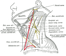
 Side of neck, showing chief surface markings. (Nerves are yellow, arteries are red.) | |
| Details | |
| Identifiers | |
| Latin | trigonum cervicale posterius trigonum colli posterius regio cervicalis posterior |
| TA98 | A01.2.02.009 |
| TA2 | 239 |
| FMA | 57778 |
| Anatomical terminology | |
The posterior triangle has the following boundaries:[1]
Apex: Union of the sternocleidomastoid and the trapezius muscles at the superior nuchal line of the occipital bone
Anteriorly: Posterior border of the sternocleidomastoideus
Posteriorly: Anterior border of the trapezius
Inferiorly: Middle one third of the clavicle
Roof: Investing layer of the deep cervical fascia
Floor: (From superior to inferior)
1) M. semispinalis capitis
2) M. splenius capitis
3) M. levator scapulae
4) M. scalenus posterior
5) M. scalenus medius
The posterior triangle is crossed, about 2.5 cm above the clavicle, by the inferior belly of the omohyoid muscle, which divides the space into two triangles:
A) Nerves and plexuses:
B) Vessels:
C) Lymph nodes:
D) Muscles:
The accessory nerve (CN XI) is particularly vulnerable to damage during lymph node biopsy. Damage results in an inability to shrug the shoulders or raise the arm above the head, particularly due to compromised trapezius muscle innervation.
The external jugular vein's superficial location within the posterior triangle also makes it vulnerable to injury.
Seamless Wikipedia browsing. On steroids.
Every time you click a link to Wikipedia, Wiktionary or Wikiquote in your browser's search results, it will show the modern Wikiwand interface.
Wikiwand extension is a five stars, simple, with minimum permission required to keep your browsing private, safe and transparent.