Tyrosinase
Enzyme for controlling the production of melanin From Wikipedia, the free encyclopedia
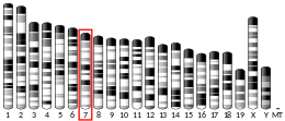
Tyrosinase is an oxidase that is the rate-limiting enzyme for controlling the production of melanin. The enzyme is mainly involved in two distinct reactions of melanin synthesis otherwise known as the Raper–Mason pathway. Firstly, the hydroxylation of a monophenol and secondly, the conversion of an o-diphenol to the corresponding o-quinone. o-Quinone undergoes several reactions to eventually form melanin.[5] Tyrosinase is a copper-containing enzyme present in plant and animal tissues that catalyzes the production of melanin and other pigments from tyrosine by oxidation. It is found inside melanosomes which are synthesized in the skin melanocytes. In humans, the tyrosinase enzyme is encoded by the TYR gene.[6]
| TYR | |||||||||||||||||||||||||||||||||||||||||||||||||||
|---|---|---|---|---|---|---|---|---|---|---|---|---|---|---|---|---|---|---|---|---|---|---|---|---|---|---|---|---|---|---|---|---|---|---|---|---|---|---|---|---|---|---|---|---|---|---|---|---|---|---|---|
| Identifiers | |||||||||||||||||||||||||||||||||||||||||||||||||||
| Aliases | TYR, ATN, CMM8, OCA1, OCA1A, OCAIA, SHEP3, tyrosinase, Tyrosinase | ||||||||||||||||||||||||||||||||||||||||||||||||||
| External IDs | OMIM: 606933; MGI: 98880; HomoloGene: 30969; GeneCards: TYR; OMA:TYR - orthologs | ||||||||||||||||||||||||||||||||||||||||||||||||||
| |||||||||||||||||||||||||||||||||||||||||||||||||||
| |||||||||||||||||||||||||||||||||||||||||||||||||||
| |||||||||||||||||||||||||||||||||||||||||||||||||||
| |||||||||||||||||||||||||||||||||||||||||||||||||||
| |||||||||||||||||||||||||||||||||||||||||||||||||||
| Wikidata | |||||||||||||||||||||||||||||||||||||||||||||||||||
| |||||||||||||||||||||||||||||||||||||||||||||||||||
Catalyzed reaction
Summarize
Perspective
Tyrosinase carries out the oxidation of phenols such as tyrosine and dopamine using dioxygen (O2). In the presence of catechol, benzoquinone is formed (see reaction below). Hydrogens removed from catechol combine with oxygen to form water.
The substrate specificity becomes dramatically restricted in mammalian tyrosinase which uses only L-form of tyrosine or DOPA as substrates, and has restricted requirement for L-DOPA as cofactor.[7]
Active site

| monophenol monooxygenase | |||||||||
|---|---|---|---|---|---|---|---|---|---|
 Catechol-Quinone | |||||||||
| Identifiers | |||||||||
| EC no. | 1.14.18.1 | ||||||||
| CAS no. | 9002-10-2 | ||||||||
| Databases | |||||||||
| IntEnz | IntEnz view | ||||||||
| BRENDA | BRENDA entry | ||||||||
| ExPASy | NiceZyme view | ||||||||
| KEGG | KEGG entry | ||||||||
| MetaCyc | metabolic pathway | ||||||||
| PRIAM | profile | ||||||||
| PDB structures | RCSB PDB PDBe PDBsum | ||||||||
| Gene Ontology | AmiGO / QuickGO | ||||||||
| |||||||||
The two copper atoms within the active site of tyrosinase enzymes interact with dioxygen to form a highly reactive chemical intermediate that then oxidizes the substrate. The activity of tyrosinase is similar to catechol oxidase, a related class of copper oxidase. Tyrosinases and catechol oxidases are collectively termed polyphenol oxidases.
Structure
Summarize
Perspective
Tyrosinases have been isolated and studied from a wide variety of plant, animal, and fungal species. Tyrosinases from different species are diverse in terms of their structural properties, tissue distribution, and cellular location.[9] No common tyrosinase protein structure occurring across all species has been found.[10] The enzymes found in plant, animal, and fungal tissue frequently differ with respect to their primary structure, size, glycosylation pattern, and activation characteristics. However, all tyrosinases have in common a binuclear, type 3 copper centre within their active sites. Here, two copper atoms are each coordinated with three histidine residues.

Plant
In vivo, plant PPOs are expressed as about 64–68 kDa proteins consisting of three domains: a chloroplastic transit peptide (containing a ~4-9 kDa thylakoid signal peptide), a catalytically active domain (~ 37–42 kDa) containing the dinuclear copper center, and a C-terminal domain (~15–19 kDa) shielding the active site.[11]
Mammalian
Mammalian tyrosinase is a single membrane-spanning transmembrane protein.[12] In humans, tyrosinase is sorted into melanosomes[13] and the catalytically active domain of the protein resides within melanosomes. Only a small, enzymatically inessential part of the protein extends into the cytoplasm of the melanocyte.
As opposed to fungal tyrosinase, human tyrosinase is a membrane-bound glycoprotein and has 13% carbohydrate content.[14]
The derived TYR allele (rs2733832) is associated with lighter skin pigmentation in human populations. It is most common in Europe, but is also found at lower, moderate frequencies in Central Asia, the Middle East, North Africa, and among the San and Mbuti Pygmies.[15]
Bacterial
In peatlands, bacterial tyrosinases are proposed to act as key regulators of carbon storage by removing phenolic compounds, which inhibit the degradation of organic carbon.[16]
Fungal
In the fungus Neurospora crassa, four different forms of tyrosinase were distinguished among different strains.[17] In each strain only one structure-determining genetic region was found for the enzyme.
Gene regulation
The gene for tyrosinase is regulated by the microphthalmia-associated transcription factor (MITF).[18][19]


Clinical significance
A mutation in the tyrosinase gene resulting in impaired tyrosinase production leads to type I oculocutaneous albinism, a hereditary disorder that affects one in every 20,000 people.[21]
Tyrosinase activity is very important. If uncontrolled during the synthesis of melanin, it results in increased melanin synthesis. Decreasing tyrosinase activity has been targeted for the improvement or prevention of conditions related to the hyperpigmentation of the skin, such as melasma and age spots.[22]
Several polyphenols, including flavonoids or stilbenoid, substrate analogues, free radical scavengers, and copper chelators, have been known to inhibit tyrosinase.[23] Henceforth, the medical and cosmetic industries are focusing research on tyrosinase inhibitors to treat skin disorders.[5]
Inhibitors
Known Tyrosinase inhibitors are the following:[24]
Genetics
Summarize
Perspective
While albinism is common, there have only been a few studies about the genetic mutations in the tyrosinase genes of animals. One of them was on Bubalus bubalis (water buffalo). The tyrosinase mRNA sequence of the wild-type B. bubalis is 1,958 base pairs (bp) with an open reading frame (ORF) of 1,593 bp long, which translates to 530 amino acids. Meanwhile, the tyrosinase gene of the albino B. bubalis (GenBank JN_887463) is truncated at position 477, caused by a point mutation in nucleotide 1431 which converts a Tryptophan (TGG) into a stop codon (TGA), resulting in a shorter and inactive tyrosinase gene.[25] Other albinos have point mutations that appear to inactivate Tyrosinase without truncation (see table and figure for examples).
| Species | Common name | Amino Acid mutation | GenBank | Uniprot ID |
|---|---|---|---|---|
| Bubalus bubalis | Water Buffalo | W477 -> Stop codon | JN_887462 | J7FBF2 |
| Pelophylax nigromaculatus | Pond Frog | Deletion of a K228 | Q04604 | |
| Glandirana rugosa | Wrinkled Frog | G376 -> D376 | A0A1I9FZH0 | |
| Fejervarya kawamurai | Rice Frog | G57 -> R57 | A0A1E1G7U0 |

Knowing that there are a few studies about the genomic data of the tyrosinase gene, there are only a handful of studies on the mutations in albino amphibians. Miura et al. (2018) investigates the amino acid mutations in the tyrosinase gene in three albino frogs: Pelophylax nigromaculatus (pond frog), Glandirana rugosa (wrinkled frog) and Fejervarya kawamurai (rice frog). In total, five different populations were studied of which three were P. nigromaculatus and one each of G. rugosa and F. kawamurai. In two of the three P. nigromaculatus populations, there was a frameshift mutation because of the insertion of a thymine within exons 1 and 3, and the third population lacked three nucleotides that encoded a Lysine in exon 1. The population of G. rugosa had a missense mutation where there was an amino acid substitution from a Glycine to Aspartic acid, and the mutation of F. kawamurai was also an amino acid substitution from Glycine to Arginine. The mutation for G. rugosa and F. kawamurai occurs in exons 1 and 3. The mutations of the third population of P. nigromaculatus, and the mutations of G. rugosa and F. kawamurai occurred in areas that are highly conserved among vertebrates which could result in a dysfunctional tyrosinase gene.[26]


Evolution
Summarize
Perspective

Tyrosinase is a highly conserved protein in animals and apparently arose already in bacteria. The tyrosinase related protein (Tyrp1) and dopachrome tautomerase (Dtc), which encode for protein implicated in melanin synthesis which are the common regulatory elements of exon/intron structure. The development of the three types of vertebrate pigment cells, although different, thus converge at a certain point to allow the expression of members of the tyrosinase family, in order to produce melanin pigments.[28] Tyrosinase family related genes plays an important role in the evolution, genetics, and developmental biology of pigment cells, as well as to approach human disorders associated with defects in their synthesis, regulation or function in vertebrates three types of melanin producing pigment cells are well known since embryonic origin i.e., from the neural crest, neural tube and pineal body. All of them have the capacity to produce melanin pigments. Their biosynthesis is governed by evolutionary conserved enzymes of the tyrosinase family( tyr, tyr1 and tyr2) also called DOPAchrome tautomerase (dct). Among them Tyr plays significance role in melanin production. However, sequenced genome from the different taxa for evolutionary analysis in the depth become more crucial in present study.[29] Similarly, the type-3 copper protein family perform various biological function including pigment formation, innate immunity and oxygen transport. The combine genetic phylogenetic and structural analysis concluded that the original type-3 copper protein possessed a single peptide and grouped into α subclass. The ancestral protein gene underwent to two duplication i.e., first one prior to divergence of unknown eukaryotic lineage and second one before diversification. The prior duplication gave rise to cytosolic form(β) and latter duplication gave membrane bound form (Γ). The structural comparison concluded that active site of α and γ forms are covered by aliphatic amino acids and β form covered with aromatic residue. Thus, the evolution of these gene family is the lineage of multicellular eukaryotes due to loss of one or more of these three subclasses and lineage-specific expansion of one or both of the remaining subclasses.[30] The genomic conserved nucleotide alignments of the tyrosinase among the vertebrate family like frogs, snakes and human suggests that it has evolved from one ancestral tyrosinase gene. The duplication and mutation of this gene is probably responsible for the emergence of a tyrosinase-related gene.[31]
Applications
Summarize
Perspective
In the food industry
In the food industry, tyrosinase inhibition is desired as tyrosinase catalyzes the oxidation of phenolic compounds found in fruits and vegetables into quinones, which gives an undesirable taste and color and also decreases the availability of certain essential amino acids as well as the digestibility of the products. As such, highly effective tyrosinase inhibitors are also needed in agriculture and the food industry.[14] Well known tyrosinase inhibitors include kojic acid,[32] tropolone,[33] coumarins,[34] vanillic acid, vanillin, and vanillic alcohol.[35]
In the cosmetic industry
Lighter skin complexion has been associated with youth and beauty across various Asian cultures. Recent research by cosmetic companies has been focused on the development of novel whitening agents that selectively suppress tyrosinase activity to reduce hyperpigmentation while avoiding cytotoxicity of healthy melanocytes.[36] Traditional pharmacological agents such as corticosteroids, hydroquinone, and amino numeric chloride lighten skin through the inhibition of melanocyte maturation.[37] However, these agents are associated with adverse effects. Cosmetic companies have been focused on developing novel whitening agents that selectively suppress the activity of tyrosinase to reduce hyperpigmentation while avoiding melanocyte cytotoxicity as tyrosinase is the rate-limiting step of the melanogenesis pathway.
In insects
Tyrosinase has a wide range of functions in insects, including wound healing, sclerotization, melanin synthesis and parasite encapsulation. As a result, it is an important enzyme as it is the defensive mechanism of insects. Some insecticides are aimed to inhibit tyrosinase.[14]
In mussel-glue inspired polymers
Tyrosinase activated polymerization of peptides, containing cysteine and tyrosine residues, lead to mussel-glue inspired polymers. The tyrosine residues are enzymatically oxidized to dopaquinones, to which thiols of cysteine could link by an intermolecular Michael-addition. The resulting polymers adsorb strongly to various surfaces with high adhesion energies.[38][39]
References
External links
Wikiwand - on
Seamless Wikipedia browsing. On steroids.


