File:Anatomy_of_the_Human_Ear_blank.svg
From Wikipedia, the free encyclopedia

Size of this PNG preview of this SVG file: 659 × 518 pixels. Other resolutions: 305 × 240 pixels | 611 × 480 pixels | 977 × 768 pixels | 1,280 × 1,006 pixels | 2,560 × 2,012 pixels.
Original file (SVG file, nominally 659 × 518 pixels, file size: 59 KB)
| This is a file from the Wikimedia Commons. Information from its description page there is shown below. Commons is a freely licensed media file repository. You can help. |
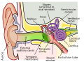
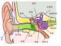
| DescriptionAnatomy of the Human Ear blank.svg |
English: A diagram of the anatomy of the human ear.
Legend: edit Polski: Schemat budowy ucha ludzkiego. |
| Date | (UTC) |
| Source | |
| Author |
|
| Other versions |
[edit]
|


| This is a retouched picture, which means that it has been digitally altered from its original version. Modifications: Deleted description. The original can be viewed here: Anatomy of the Human Ear.svg:
|
I, the copyright holder of this work, hereby publish it under the following license:
This file is licensed under the Creative Commons Attribution 2.5 Generic license.
- You are free:
- to share – to copy, distribute and transmit the work
- to remix – to adapt the work
- Under the following conditions:
- attribution – You must give appropriate credit, provide a link to the license, and indicate if changes were made. You may do so in any reasonable manner, but not in any way that suggests the licensor endorses you or your use.
Original upload log
This image is a derivative work of the following images:
- File:Anatomy_of_the_Human_Ear.svg licensed with Cc-by-2.5
- 2009-04-28T21:49:54Z Mike.lifeguard 800x600 (91972 Bytes) Malleus and incus were swapped O.o
- 2009-02-15T14:59:07Z Inductiveload 800x600 (91617 Bytes) added tympanic cavity
- 2009-02-15T14:50:03Z Inductiveload 800x600 (89435 Bytes) {{Information |Description={{en|1=A diagram of the anatomy of the human ear.}} |Source=[[:File:10.1371_journal.pbio.0030137.g001-L.jpg]], vectorised by [[User:Inductiveload|Inductiveload]] |Author=[[User:Inductiveload|Induct
Uploaded with derivativeFX
Captions
A cross-sectional diagram of the human ear.
A cross-sectional diagram of the human ear.
Items portrayed in this file
depicts
16 September 2009
image/svg+xml
fdf89a74fba97eef49b25e707979535143acded7
60,794 byte
518 pixel
659 pixel
File history
Click on a date/time to view the file as it appeared at that time.
| Date/Time | Thumbnail | Dimensions | User | Comment | |
|---|---|---|---|---|---|
| current | 16:53, 12 April 2019 | | 659 × 518 (59 KB) | Mikael Häggström | Removed misleading green area: The pinna is also part of the outer ear |
| 15:42, 10 September 2018 |  | 659 × 518 (61 KB) | Jmarchn | Bigger (proportional real size) and full redraw (more realistic) of the auricle. Ossicles in white colour. Eardrum with contour. Added 3 labels. Add fundus to the bone and subcutaneous tissues, add superior auricular muscle, add transparency to middle ear, add separation between middle and inner ear, add division to internal auditory canal. | |
| 12:15, 16 September 2009 |  | 730 × 556 (71 KB) | M.Komorniczak | {{Information |Description={{en|1=A diagram of the anatomy of the human ear.}} {{pl|Schemat budowy ucha ludzkiego.}} |Source=*File:Anatomy_of_the_Human_Ear.svg |Date=2009-09-16 12:14 (UTC) |Author=*File:Anatomy_of_the_Human_Ear.svg: Chittka L, |
File usage
No pages on the English Wikipedia use this file (pages on other projects are not listed).
Global file usage
The following other wikis use this file:
- Usage on ca.wiktionary.org
- Usage on meta.wikimedia.org
- Usage on pl.wikipedia.org
- Kosteczki słuchowe
- Młoteczek
- Strzemiączko
- Ucho
- Szablon:Ucho
- Trąbka słuchowa
- Kowadełko
- Błona bębenkowa
- Ślimak (anatomia)
- Ucho zewnętrzne
- Kanały półkoliste
- Małżowina uszna
- Przewód słuchowy zewnętrzny
- Narząd Cortiego
- Śródchłonka
- Błędnik błoniasty
- Ucho środkowe
- Jama bębenkowa
- Błędnik kostny
- Przychłonka
- Łagiewka (anatomia)
- Woreczek
- Mięsień napinacz błony bębenkowej
- Mięsień strzemiączkowy
- Musculus fixator stapedis
- Staw kowadełkowo-młoteczkowy
- Schody przedsionka
- Schody bębenka
- Jama sutkowa
- Przewody półkoliste
- Wodociąg przedsionka
- Przedsionek ucha wewnętrznego
- Przewód śródchłonki
- Worek śródchłonki
- Płatek ludzkiej małżowiny usznej
- Okienko przedsionka
- Okienko ślimaka
- Wikipedysta:Paweł Ziemian/Navi/Ucho
- Wikipedysta:Paweł Ziemian/Navi/Ucho2
- Usage on pl.wiktionary.org
- Usage on rm.wikipedia.org
Metadata
This file contains additional information, probably added from the digital camera or scanner used to create or digitize it.
If the file has been modified from its original state, some details may not fully reflect the modified file.
| Width | 658.57141 |
|---|---|
| Height | 517.71429 |