オトガイ孔
ウィキペディアから


位置・形態
オトガイ孔は通常成長と共に後方に移動し、成人では半数以上が下顎第二小臼歯の位置にある[2]。
無歯顎や大臼歯・小臼歯欠損の人ではオトガイ孔の位置は低くなる[3][4]。
通常、オトガイ孔は左右1対であるが、3.5%から24.6%の割合で複数のオトガイ孔が認められる事があり、通常大きさは揃っておらず一つの大きなオトガイ孔とその他の副オトガイ孔となる[5]。左右とも副オトガイ孔を認めるものは少ない[5]。

関連画像
 Side view of the skull.
 The skull from the front.
 Distribution of the maxillary and mandibular nerves, and the submaxillary ganglion.
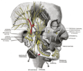 Mandibular division of the trifacial nerve.
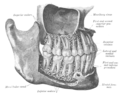 The permanent teeth, viewed from the right.


脚注
外部リンク
Wikiwand - on
Seamless Wikipedia browsing. On steroids.