File:Anatomy_of_Human_Ear_with_Cochlear_Frequency_Mapping.svg
Da Wikipedia, l'enciclopedia encyclopedia

Dimensioni di questa anteprima PNG per questo file SVG: 674 × 519 pixel. Altre risoluzioni: 312 × 240 pixel | 624 × 480 pixel | 998 × 768 pixel | 1 280 × 986 pixel | 2 560 × 1 971 pixel.
File originale (file in formato SVG, dimensioni nominali 674 × 519 pixel, dimensione del file: 33 KB)
| Questo file e la sua pagina di descrizione (discussione · modifica) si trovano su Wikimedia Commons (?) |
Dettagli
| DescrizioneAnatomy of Human Ear with Cochlear Frequency Mapping.svg |
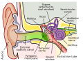
English: The human ear and frequency mapping in the cochlea. The three ossicles incus, malleus, and stapes transmit airborne vibration from the tympanic membrane to the oval window at the base of the cochlea. Because of the mechanical properties of the basilar membrane within the snail-shaped cochlea, high frequencies will produce a vibration peak near the oval window, whereas low frequencies will stimulate receptors near the apex of the cochlea (locations for three frequencies indicated schematically). Information from the cochlear receptor cells is transmitted to the cochlear nuclei via the 8th cranial nerve, and on through the midbrain to the cortex. |
| Data | |
| Fonte | Opera propria (Testo originale: Own work by uploader, derived from File:Anatomy_of_the_Human_Ear.svg ) |
| Autore | Inductiveload |
| Licenza (Riusare questo file) |
Questo file è disponibile in base alla licenza Creative Commons Attribuzione-Condividi allo stesso modo 2.5 Generico
|
| Altre versioni |
[modifica]
|
| SVG sviluppo InfoField | Questa grafica vettoriale è stata creata con Inkscape. This file is translated using SVG switch elements: all translations are stored in the same file. |


Didascalie
Aggiungi una brevissima spiegazione di ciò che questo file rappresenta
A labelled cross-sectional diagram of the human ear.
A labelled cross-sectional diagram of the human ear.
15 feb 2009
image/svg+xml
4c58b13c29ef1eb6b3ba8c18aed2cd55519d51d1
34 114 byte
519 pixel
674 pixel
Cronologia del file
Fare clic su un gruppo data/ora per vedere il file come si presentava nel momento indicato.
| Data/Ora | Miniatura | Dimensioni | Utente | Commento | |
|---|---|---|---|---|---|
| attuale | 23:29, 16 set 2018 | | 674 × 519 (33 KB) | JoKalliauer | added systemLanguage="eo" |
| 19:21, 16 set 2018 |  | 674 × 519 (32 KB) | JoKalliauer | added systemLanguage="de" | |
| 07:33, 11 set 2018 |  | 674 × 519 (87 KB) | Jmarchn | Bigger (proportional real size) and full redraw (more realistic) of the auricle. Ossicles in white colour. Eardrum with contour. Added 3 labels. Add fundus to the bone and subcutaneous tissues, add superior auricular muscle, add transparency to middle ear, add separation between middle and inner ear, add division to internal auditory canal. | |
| 15:40, 29 apr 2009 |  | 800 × 600 (98 KB) | Inductiveload | swap incus/malleus | |
| 17:10, 15 feb 2009 |  | 800 × 600 (98 KB) | Inductiveload | {{Information |Description={{en|1=The human ear and frequency mapping in the cochlea. The three ossicles incus, malleus, and stapes transmit airborne vibration from the tympanic membrane to the oval window at the base of the cochlea. Because of the mechan |
Pagine che usano questo file
Nessuna pagina utilizza questo file.
Utilizzo globale del file
Anche i seguenti wiki usano questo file:
- Usato nelle seguenti pagine di en.wikipedia.org:
- Usato nelle seguenti pagine di en.wikibooks.org:
- Usato nelle seguenti pagine di eo.wikipedia.org:
- Usato nelle seguenti pagine di he.wikipedia.org:
- Usato nelle seguenti pagine di incubator.wikimedia.org:
- Usato nelle seguenti pagine di lt.wikipedia.org:
- Usato nelle seguenti pagine di meta.wikimedia.org:
- Usato nelle seguenti pagine di www.wikidata.org:
Metadati
Questo file contiene informazioni aggiuntive, probabilmente aggiunte dalla fotocamera o dallo scanner usati per crearlo o digitalizzarlo. Se il file è stato modificato, alcuni dettagli potrebbero non corrispondere alla realtà.
| Larghezza | 674.23 |
|---|---|
| Altezza | 518.84 |