The colorectal adenoma is a benign glandular tumor of the colon and the rectum. It is a precursor lesion of the colorectal adenocarcinoma (colon cancer).[1][2][3] They often manifest as colorectal polyps.
| Colorectal adenoma | |
|---|---|
 | |
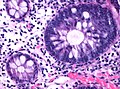
| Tubulovillous adenoma (tubular component – left of image, villous component – right of image). H&E stain. | |
| Specialty | Gastroenterology |
| Symptoms | Asymptomatic, rectal bleeding |
| Complications | Colorectal cancer |
| Diagnostic method | Colonoscopy |
| Treatment | Polypectomy |
Comparison table

| Type | Risk of containing malignant cells | Histopathology definition |
|---|---|---|
| Tubular adenoma | 2% at 1.5 cm[4] | Over 75% of volume has tubular appearance.[5] |
| Tubulovillous adenoma | 20% to 25%[6] | 25–75% villous[5] |
| Villous adenoma | 15%[7] to 40%[6] | Over 75% villous[5] |
| Sessile serrated adenoma (SSA)[8] |
|
Tubular adenoma
In contrast to hyperplastic polyps, these display dysplasia.[citation needed]
Tubulovillous adenoma
Tubulovillous adenoma, TVA are considered to have a higher risk of becoming malignant (cancerous) than tubular adenomas.[9]
 Normal (left) versus dysplastic (large at right) colonic crypts, the latter conferring a diagnosis of a tubular and/or villous adenoma.
 Histopathology of high-grade dysplasia in a tubulovillous adenoma, in this case seen mainly as loss of cell polarity, as cells become more plump and haphazard than the elongated and parallel nuclei of surrounding low-grade dysplasia.


Villous adenoma

These adenomas may become malignant (cancerous). Villous adenomas have been demonstrated to contain malignant portions in about 15–25% of cases, approaching 40% in those over 4 cm in diameter.[7] Colonic resection may be required for large lesions. These can also lead to secretory diarrhea with large volume liquid stools with few formed elements. They are commonly described as secreting large amounts of mucus, resulting in hypokalaemia in patients. On endoscopy, a "cauliflower' like mass is described due to villi stretching. Being an adenoma, the mass is covered in columnar epithelial cells.[citation needed]
Sessile serrated adenoma

Sessile serrated adenomas are characterized by (1) basal dilation of the crypts, (2) basal crypt serration, (3) crypts that run horizontal to the basement membrane (horizontal crypts), and (4) crypt branching. The most common of these features is basal dilation of the crypts.
See also
References
Wikiwand in your browser!
Seamless Wikipedia browsing. On steroids.
Every time you click a link to Wikipedia, Wiktionary or Wikiquote in your browser's search results, it will show the modern Wikiwand interface.
Wikiwand extension is a five stars, simple, with minimum permission required to keep your browsing private, safe and transparent.