Loading AI tools
| This It is of interest to the following WikiProjects: | ||||||||||||||||||
| ||||||||||||||||||
Is there any reason the image isn't showing up on this page? The larger version doesn't seem to be working either. Might be I'm just missing something? -- TomPreuss 13:34, 5 Aug 2004 (UTC)
- I don't think there's anything wrong with the formatting of the page, it'd show the image if the image existed - I get a 404 not found message when I try to look at the image directly. I guess the image needs to be uploaded to Wikipedia again. Average Earthman 15:59, 5 Aug 2004 (UTC)
- That's pretty odd though. What do you think could cause that to happen? -- TomPreuss 13:03, 6 Aug 2004 (UTC)
the work is perfect if i may add — Preceding unsigned comment added by 129.122.0.121 (talk) 12:03, 7 February 2022 (UTC)
Shouldn't this page be listed under SEM? It's a pretty common abbreviation. --Michaeltoe 22:09, 4 Feb 2005 (UTC)
The use of a dedicated backscattered electron detector above the sample in a "doughnut" type arrangement, with the
That is all: the paragraph in the section 'Detection of backscattered electrons' ends abruptly. Seems like there's some half-editing... I can now see the paragraph fully, and no one has edited it!! Perhaps there was a problem in the browser I was using(in my college). Firefox proves itself to be THE best. :)
SundarKanna 20:03, 26 December 2005 (UTC)
- The rest of that paragraph had lost in a revert some time ago. I put the missing section back in yesterday. eaolson 21:08, 26 December 2005 (UTC)
I removed the template {{FAOL|French|fr:Microscopie électronique à balayage}} because the French article lost its featured article status. Stefane 07:47, 21 March 2006 (UTC)
What are secondary electrons? What does "escape" mean? Why do they "escape"? This section does not stand on its own, but relies on a careful reading of other sections. While I made some changes due to style and grammar in this section, I am still confused by its content. I don't want to change the article itself because of my lack of knowledge, but here is what I think it is trying to say. I have heavily interpreted what I read and even made educated guesses. Can anyone confirm/disconfirm my guesswork?
"An electron beam rasters (i.e. scans back and forth) over the surface of a material, which absorbs the energy. The material re-emits secondary electrons in every direction. An electron sensor known as a scintillator-photomultiplier, measures the intensity of the secondary electrons and converts it into a two-dimensional image with bright and dim regions. The most common imaging technique detects low energy (<50 eV) secondary electrons originating a few nanometers from the surface.
The brightness depends on the angle of the surface. If the beam is perpendicular to the surface, then the spotlighted region is as small as possible, which minimizes the number of electrons that escape. As the surface angle increases, the spotlighted area increases, and more secondary electrons will be emitted. Steep surfaces and edges appear brighter than flat surfaces, resulting in well-defined, three-dimensional images. This technique makes resolutions of less than 1 nm possible." — Preceding unsigned comment added by Dhskep (talk • contribs) 05:11, 6 April 2006 (UTC)
- There surely is an error in the section as it is writing in the post right now; "As the angle of incidence increases, the "escape" distance of one side of the beam will decrease, and more secondary electrons will be emitted."
- This "ecape distance" must increase with angle and secondary atoms emitted.
- But you version without escape distance is certainly much more eatable then the current. —Preceding unsigned comment added by Nanopete (talk • contribs) 22:16, August 25, 2007 (UTC)
What are secondary electrons? What does "escape" mean? Why do they "escape"?
— User:Dhskep
Secondary electrons are inelastically scattered electrons of low energy. Primary electrons interact with an electron in an atom of the material and kick it out, leaving a hole. Depending on where it is in the atom, this hole can lead to an xray emission, auger electron, blah blah. The primary electron loses energy in the interaction, then generally goes on its merry way. The ejected electron is the secondary, and it has very low energy, i.e. it is moving very slowly. There is nothing particular to the surface that is conducive to the production of secondaries, but because they move slowly, they can only escape from near the surface. They have a very short mean free path. Detection is done by attracting secondaries with (usually) two bias potentials. A large wire cage over the SE detector will have a variable bias, usually +/-300V to attract or repel SEs. Behind that is a scintillator which will be biased at 8-10kV. Secondary electrons are attracted to the cage, then the scintillator.
There can be additional complicating factors with SEs. Because the scintillator has line of sight exposure to the specimen, it also inadvertently detects BSEs. Additionally, SEs fit in two categories. SE1, generated by the impact of the primary beam, and SE2, generated by escaping BSEs. The SE1/SE2 problem can be mitigated by using lower accelerating voltages and the inadvertent BSEs can be mitigated by using filtered detectors (eg. Hitachi ExB, Zeiss ESB, JEOL r-filter, FEI yadayada). Those filtered detectors are only available on FESEMs, probably for market segmentation and cost reasons. --137.82.22.226 (talk) 16:52, 3 December 2019 (UTC)
I came across an orphan about OIM (apparently stands for orientation imaging microscopy). Can some one try to verify the information in it? Here: Orientation imaging microscopy (which is where I had to move it to make way for an "OIM" disambiguation page. — User:Donama 04:33, 17 May 2006 (UTC)
- Fixed. 24.211.234.99 22:30, 15 October 2006 (UTC)
Can somebody give me the construction and the electronics involved in electron microscopy? —Preceding unsigned comment added by 220.225.87.34 (talk • contribs) 06:25, 11 June 2007 (UTC)
- It consists of an electron gun commonly made of tungsten and cathode because of its high melting point.a grid is placed nearer to the electron gun which is used to accelerate the primary eletrons.a magnetic condensor lens and scanning coils is placed nearer to the grid .sample is placed next to the scanning coil a collector is followed by a video amplifier and cro .the scanning coil is connected to the cro and it is then connected to the scan generator. —Preceding unsigned comment added by 117.193.98.168 (talk • contribs) 02:19, 17 November 2009 (UTC)
The reference to Alan Nelson's patent is irrelevant as there has never been any implementation of this approach to an environmental SEM either in laboratory equipment
or in commercial instruments, and no published reference to a refereed journal exists. In contrast, the patent:
U.S. Patent No. 4,823,006, filed Feb. 19, 1988. Integrated electron optical/differential pumping imaging signal detection system for an environmental scanning electron microscope. Inventors: G.D. Danilatos, G.C. Lewis. Assignee: ElectroScan Corporation.
describes the first commercial ESEM imlementation by ElectroScan. The latter was based on prior Danilatos works as listed in http://www.danilatos.com. In addition to the differential pumping and electron detection techniques described by the first Danilatos reference provided in the present entry, the commercial ESEM has made extensive use of the newly invented system of a gaseous detection device as comprehensively described by:
Danilatos, G.D. (1990). Theory of the Gaseous Detector Device in the ESEM in Advances in Electronics and Electron Physics, Academic Press, Vol. 78:1-102.
The Alan Nelson patent inappropriately employs a series of apertures with trapped air in the cavities between them. As a result, the electron beam would suffer significant losses before it can be usefully employed in the specimen chamber. At any rate, Alan Nelson by no means should be referenced as a contributor to the development of ESEM, let alone as an initiator of this technology. In contrast, Danilatos preceded with his first patent and papers on ESEM in 1979. It is therefore proposed to amend the references accordingly.
Esem0 05:40, 8 July 2007 (UTC) Esem0 05:43, 8 July 2007 (UTC) Esem0 05:45, 8 July 2007 (UTC)
Esem0 04:11, 10 July 2007 (UTC) Esem0 04:12, 10 July 2007 (UTC)
I've been looking through some of the imaging articles, and many appear to be missing major concepts fundamental to the instrument. For example, SEMs are known for their amazing depth of field, yet this term is not used at all in this article. Is there something I don't know about SEMs or Wikipedia and imaging articles that is a reason for omitting this?
KP Botany 02:06, 4 October 2007 (UTC)
it would be very nice if resource for downloadable SEM images On an archived bundle (such as rar or zip) also put here as the resource for people yo get Free SEM images on a specific category, such as human cells or parasitology Etc.
ive been looking anywhere to get such resource, and nothing better than here. But i wish a can find The free image bundled as i mention. —Preceding unsigned comment added by Kuplukjaya (talk • contribs) 07:06, 25 March 2008 (UTC) Kuplukjaya (talk) 07:13, 25 March 2008 (UTC)
This edit changed a fair bit of text, and seems to have removed some info, as well as adding some. Can someone look at this? Should it simply be reverted? —Pengo 01:11, 30 March 2008 (UTC)
There are many poor quality SEM images on Wikipedia for some reason. I removed some from the gallery for this article, because there are a sufficient number of better quality or useful images. The one of the topo/atomic number contrast should also be removed when a better image comes up.
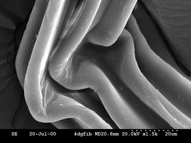
The first one I removed is of the polyester. This image has serious charging artifacts, creating the ruinous line across the middle of the image, it's in focus almost nowhere, and it has lousy depth of field (of course, since it's in focus almost nowhere). A longer working distance, a smaller aperture, a smaller probe size, lower KV, better coating, many things would have made this a better image. It's fine for the polyester fiber article for now, but a better one should be captured or found, as this one has too many problems with it to be used in an encyclopedia for anything other than an article on SEM artifacts (which it would be good for).
The diatoms are out of focus. There's no point to the image. It's not in focus anywhere in the depth of focus of the image.
The red blood cells should be removed also, when they can be replaced with a better image. Red blood cells are easy to shoot on an SEM, and this image is far too noisy to be considered for production anywhere outside of Wikipedia--it's too old. However, because red blood cells look totally cool on SEM (versus TEM), it's okay to leave this very noisy image in for now. They're a standard.
The nematocyst is too low resolution to be of any use. It should be replaced, also.
The Drosophila melanogaster body is very poorly preserved, rather than life-like, there's excessive charging to the point of putting a bright white lightening bolt through the image, and the carbon dag substrate consumes an unwieldy portion of the image. However, this is a basic subject in SEM, like red blood cells. Its compound eye is completely out of focus, also.
The asbestos fibers should be photoshopped to remove the charging artifacts, if left in. The picture is not compelling or illustrative of anything other than the fact that SEM is used to look at asbestos fibres. I think it is rather common.
Probably the same public domain sources could be searched for better images.
An SEM image should be in focus. This does not require an expert microscopist to discern. It's either in focus or it's not. TEM images can be much more subjective and demanding of expert eyes to tell quality. Especially with biological specimens, if it looks out of focus, if the insect is collapsed and dessicated, it's not a good image. It's more difficult with materials specimens, but there are few of any of these of any quality on Wikipedia.
--Blechnic (talk) 06:27, 8 April 2008 (UTC)
- Then you should replace rather than remove if you are concerned about image quality. Otherwise you are depriving readers of information. Peterlewis (talk) 06:36, 8 April 2008 (UTC)
- Precisely what information have I deprived anyone of by removing an unacceptable micrograph? Can I upload pictures of my cat and use it, no matter how low quality, in the article on cats? No. The same thing should not be acceptable with micrographs. I left plenty of bad ones without leaving the worthless in the article. They contain no information without pointing out to the readers that they are useless due to artifacts which render them of too low quality to be proper micrographs. --Blechnic (talk) 06:51, 8 April 2008 (UTC)
- PS In addition, people with good micrographs may just see an overly full gallery. They won't want to add good images in with the low quality ones, or they may not even look beyond the number of micrographs, thinking Wikipedia is full of SEM images. It's not. --Blechnic (talk) 06:53, 8 April 2008 (UTC)
- Precisely what information have I deprived anyone of by removing an unacceptable micrograph? Can I upload pictures of my cat and use it, no matter how low quality, in the article on cats? No. The same thing should not be acceptable with micrographs. I left plenty of bad ones without leaving the worthless in the article. They contain no information without pointing out to the readers that they are useless due to artifacts which render them of too low quality to be proper micrographs. --Blechnic (talk) 06:51, 8 April 2008 (UTC)
- Then you should replace rather than remove if you are concerned about image quality. Otherwise you are depriving readers of information. Peterlewis (talk) 06:36, 8 April 2008 (UTC)
Additional question on images. Does someone have the original source material for the photoresist? It says it is taken at 1KV, it also has "standing waves" and "DSM" on the bottom. 1 KV is very unusual for SEM, so it should be elaborated in the caption. --Blechnic (talk) 07:01, 8 April 2008 (UTC)
- I think you are being over-critical. I am a microscopist myself, and would be happy to use any of the images you deleted. If you have better ones or more showing other details, then put them in. But you cannot just delete what is already there. Peterlewis (talk) 07:06, 8 April 2008 (UTC)
- Yes, if the images are completely unworthy due to artifacts that could not be published, they should be removed. I am skeptical that you are a microscopist and would leave an image like that of the blurry, out of focus, no depth of field polyester with all the charging and a charging artifact slicing the image almost in half top to bottom, the blown out asbestos, the flattened flies, the out of focus diatoms. Leaving junk in is an invitation not to find much better. Especially compared to the newer micrographs. There are plenty of images left in the gallery that leaving the junk in is not necessary. --Blechnic (talk) 07:10, 8 April 2008 (UTC)

This image is in focus in over less than 10% of the picture. It has no depth of field--it might as well have been shot on a light microscope. It has ugly charging artifacts. The substrate (cracked dag?) is hideous. The information on the bottom shows what went wrong: too short a depth of field, too high a KV, add the blown out areas, and the charging artifacts and you wind up with not much worth sharing. Just because it's a secondary electron micrograph doesn't mean it has to be on Wikipedia. --Blechnic (talk) 07:16, 8 April 2008 (UTC)
Well, stop moaning about images and replace the ones you object to with your own. I have been using SEM, ESEM and OM for many years and live with the problems you mention. If only we all lived in a perfect world! Perhaps you should write a new article on "artefacts in SEM". Peterlewis (talk) 08:07, 8 April 2008 (UTC)
- I'm not sure how much value it would be to a general encyclopedia. I get images all of the time with charging, edge effect, out of focus, not enough depth of field, too low in contrast. But I shoot them over if I need them to illustrate a point. I don't publish them for the whole world as an example of an electron micrograph. That's the difference, here, and it's an important one. I'm not moaning about the images. I stated precisely why I removed the ones I did, why I left the ones I did, and why the removed ones should not stay in the article. This is not "moaning," it is a courtesy to other readers to explain what is going on. I also elaborated to you when you asked. --Blechnic (talk) 09:11, 8 April 2008 (UTC)
- You should be more positive, and put some of your pics into the article before deleting the work of others. The present set (which I have restored) are quite acceptable in my opinion for the general reader. However, what you could do is to add comments about charging and lack of focus etc as a separate section, and then show them how to eliminate such defects if they are interested in the finer detail. Peterlewis (talk) 09:25, 8 April 2008 (UTC)
- So, explaining how bad they are would satisfy you? How would that enhance the article? Is that standard for Wikipedia, that you include bad pictures that poorly illustrate a point, then explain what's so bad about them? Why have so many pictures, if so many of them are bad? Well, suit yourself, I'll label them why they're garbage. --Blechnic (talk) 09:28, 8 April 2008 (UTC)
- You should be more positive, and put some of your pics into the article before deleting the work of others. The present set (which I have restored) are quite acceptable in my opinion for the general reader. However, what you could do is to add comments about charging and lack of focus etc as a separate section, and then show them how to eliminate such defects if they are interested in the finer detail. Peterlewis (talk) 09:25, 8 April 2008 (UTC)
- Okay, I labeled them as junk. I'll see what other junk I can upload to the gallery, since it seems, that having a few quality images is a bad thing when one can have a ton of images that do nothing to enhance the article, are low quality, barely show what they are supposed to show, and simply waste band width. But you're the microscopist. Silly of me to think one quality image could show as much as three junk shots. --Blechnic (talk) 09:38, 8 April 2008 (UTC)
- Look, I've stumbled across this talk page by chance after having a disagreement with you on featured picture candidates, and I can see quite clearly that you have a very poor attitude and a stubbornness that makes it difficult for you to appreciate any other point of view, looking down on those you disagree with. I'm no expert on SEM, but I do agree with Peterlewis that until better images are procured, either by yourself or others, there is no harm in keeping existing ones that illustrate unique subjects. As he mentioned, sure, comment on what could have been done better if you think it would benefit the article, but the article is not just for experts, and it is quite likely that laymen wouldn't even appreciate or care how bad the images are, technically.
- Ultimately though, why not just improve on the images rather than remove them and complain about how horrible they are? Diliff | (Talk) (Contribs) 22:32, 10 April 2008 (UTC)
My "attitude" and "stubborness?" Are we discussing the article or attacking me personally? I look down on those I disagree with? Where's your proof of that? I'm too short. There is no need to put any crappy image on Wikipedia for any reason. It's not a dumping or holding ground for garbage, particularly when the gallery can be filled nicely with a smaller but sufficient number of SEM micrographs that actually show something. If you wish to discuss me and your guesses about me personally and attack me personally find somewhere and some way else to do it. This is a discussion page for the SEM article. The article was improved by removing the images, rather than by littering it with crap by keeping the images in. The existing ones do not illustrate unique subjects. I left the ones that do this in. The article doesn't have to be for experts to show a decent SEM micrograph. That is the beauty of an SEM, even non-experts can look at a beautiful micrograph, like the ones of the arthropod eyes, and tell they are beautiful and without the artifacts of bad micrographs. Littering Wikipedia with crap in articles simply because someone uploaded it isn't writing an encyclopedia. Peter Lewis insists the crap must stay, so it should be identified as crap. In my opinion a smaller gallery of a well-chosen variety of good images would be a much better choice, but Peter Lewis wants the bad micrographs, so I agreed to allow them with the captions. How about I divide the gallery in two? Example electron micrographs and trash? Get off the topic of me. --Blechnic (talk) 04:44, 11 April 2008 (UTC)
This, right here, is a nice gallery of notable, good, or example electron micrographs:
- False coloured SEM image of soybean cyst nematode and egg.
- Compound eye of Antarctic krill Euphausia superba.
- Ommatidia of Antarctic krill eye.
- Noisy SEM image of normal circulating human blood.
- SEM image of a hederelloid from the Devonian of Michigan (largest tube diameter is 0.75 mm).
- Backscattered Electron image of an Antimony rich region in a fragment of ancient glass.
- SEM image of the corrosion layer on the surface of an ancient glass fragment, note the laminar structure of the corrosion layer.
- SEM image of a photoresist layer used in semiconductor manufacturing taken on a Field emission SEM at 1000 volts, a very low accelerating voltage for an SEM, achievable in Field Emission SEMs.
Adding garbage to the article, simply because it exists on Wikipedia, does not make this a better article, or a better gallery. It is just poor quality, unpublishable, micrographs. Why publish them on Wikipedia? Is this a dumping ground for bad snapshots? No, this is an encyclopedia article. More is not better, when the more is without additive value. Adding poor quality micrographs that fail to illustrate anything but their own poor quality is not necessary for an article on SEM. I bet the photography and camera articles don't have example trash images in their galleries. It's absurd. --Blechnic (talk) 04:51, 11 April 2008 (UTC)
- Peterlewis is right: If Blechnic knows so well what constitutes good SEM images, then certainly he should be in a position to supply them too. Instead, he just repeats the words "crap" and the like without making any step forward. Removing the images is a step backward, and I can see no harm to the reader for the purposes of this very short entry. I think Blechnic should put up or quit arguing. It's absurd, indeed. I have been working in the field for 30 years. Thank you. — Preceding unsigned comment added by Esem0 (talk • contribs) 12:01, 13 April 2008 (UTC)
- There's no need to put up or shut up anything. The gallery is a discrete, usable gallery of a variety of SEM images, each with a purpose. There's no need to litter any Wikipedia article with crap, just because crap is available, as there is no end to available crummy micrographs. I'm not impressed with you working in the industry for 30 years if you see a need to keep a poorly captured, out of focus throughout entire image, micrograph in an article. Over 30 years you should have seen thousands of far better micrographs and no need whatsoever to keep an amateurish one anywhere, much less give it prominent in an encyclopedia article. Wikipedia is the only encyclopedia featuring bad micrographs. --Blechnic (talk) 20:10, 13 April 2008 (UTC)
- I won't comment on the quality of the images, but I would say that I'd feel the aim of the gallery would be to show the whole range of capability of the SEM, rather than contain a large number of images of a similar type (e.g. secondary electron images of parts of insects). As such, a lower quality image that illustrates a different application could be preferable to a very good image of a type of which we have more than one example already. DrMikeF (talk) 10:33, 7 August 2008 (UTC)
- Re the caption on the photoresist about the semiconductor industry: the purpose of mentioning their importance in the industry is that the image is of a semiconductor photoresist. Yes, they're important (high res FE guns) in many industries, but these are just notes about the particular images in the gallery, so the high res comment is specific to this excellent micrograph. --Blechnic (talk) 20:10, 13 April 2008 (UTC)
"Gold coating may be regarded as a semi-destructive process since removal of a gold coating requires aggressive chemicals such as potassium cyanide or aqua regia, although this would only be required in practise if a specimen has high intrinsic value, as in an archaeological artefact."
Is there an example of a museum coating an archaeological artefact with "high intrinsic value" then using arsenic of something to remove the gold? I pretty much only work with natural history specimens, often type specimens, so coating is simply not done, so it's hard to imagine damaging a precious specimen by coating it to capture an image. An example would go a long way for this sentence. --Blechnic (talk) 02:40, 23 April 2008 (UTC)
- In 30 year's work with SEMs I have only very rarely encountered a requirement to uncoat a specimen. It can be done, however, though using arsenic you'll probably not get very far.Plantsurfer (talk) 08:27, 23 April 2008 (UTC)
- Then let's remove this statement entirely. It's weird. I've uncoated a few specimens, but never archaelogical museum specimens. --Blechnic (talk) 16:53, 23 April 2008 (UTC)
- Go on then, why don't you?Plantsurfer (talk) 17:02, 23 April 2008 (UTC)
Also, I thought "Environmental SEM" was a trademarked name? If so, should we be using it generically in an encyclopedia, or does this matter?
"Working with the method is easier because the sample chamber is very large, and control is usually completely computer controlled."
Always very large? I just visited a lab today with an old Zeiss SEM (tungsten filament) that has a huge sample chamber. The variable vacuum SEM I use has a pretty big sample chamber, but it's not huge compared to my other SEMs. And, "completely computer controlled" is not a function of low vacuum variable pressure SEMs, it's a function of what you ordered when you buy it. It's conceptually new so it comes with a computer interface, but the cheaper versions can come with mechanical controls if you're an institution with a finite budget and a high need.
"Coating is thus unnecessary, and X-ray analysis unhindered. Sample manipulation within the specimen chamber is always more difficult than in optical microscopy, however, and colour rendition is absent."
Huh? Why do we go to comparing an environmental SEMs chamber manipulation with that of a light microscope? Is it easier in a conventional SEM. And, no, it's not "always more difficult than in optical microscopy!" It depends upon the optical microscope and the SEM. Last week it took me all day to set up some light microscope images and half an hour to shoot a few dozen high resolution bacterial images on an SEM, mostly because of the time spent aligning the optical microscope's stage for collecting data.\
"Colour rendition is absent" in an environmental SEM? Well, why would anyone use them if they can't get color? It's also absent in all electron microscopy. This section needs to be about the ESEM, not just filled with general information to make it thicker. Maybe it was just copied from another article merged with this?
I'm beginning to think someone writing this article is selling or investing in ESEMs. However, it's not necessarily to internally link every occurrence to its section in this article. People will still buy them without the excessive linking.
--Blechnic (talk) 02:40, 23 April 2008 (UTC)
- excellent, there was a lot of POV in those sections Plantsurfer (talk) 08:27, 23 April 2008 (UTC)
Indeed, the removal of gold-coating is such a minute detail in the big picture of SEM, weird to have it here. I agree that the link of ESEM to FEI company should be deleted. They do not have the monopoly of it any more. However, the name of ElectroScan should remain as it is historically important. The names ESEM and Environmental SEM are generic terms in use for about three decades long before ElectroScan claimed it as a trademark (this term was actually first introduced by Danilatos in 1980). Therefore, the fact that a commercial company successfully but "unlawfully" managed to trademark it for some time has created a misconception which should not be allowed to leave a permanent imprint in the encyclopedia. Commercialism should be struck out. If you were to delete ESEM, then what else is there to replace it with? Perhaps, VPSEM which is used by a competitor to FEI to also promote ESEM (who use ESEM as generic term in their brochures); or "Wet-SEM" adopted yet by another competitor? Or "Natural SEM" by another one, or "Bio-SEM" once used by AMRAY? The encyclopedia should not be trapped by any on these commercialisms with their corresponding catchwords. However, ESEM is an independent generic term naturally evolved from academic research. Manufacturers have unfortunately created a lot of confusion around, and many workers in the field have been misled. It would be nice if all involved in this subject took some time to study the relevant literature. There are many misconceptions around and things should be cleared once and for all. This forum is a good place to start. Please comment before action is taken to make the change - or the first proposer (or other) proceed to make it. Esem0 (talk) 00:44, 26 April 2008 (UTC)
- Thanks for the lecture, but the question does remain. Is it a trademarked term? Or not?
- This is not really a forum, and not the place to disabuse folks of misconceptions about nomenclature usage in the field of scanning electron microscopy, it's simply the discussion page for this article.
- It is however useful to discuss and agree upon actions before making changes, like discussing a general outline for this article. It would be very useful.
- However, this is also not an article about environmental SEMs, and there was simply too much about it in the article, and the same information repeated over and over, in in appropriate places, often. I, by the way, don't call VP SEMs "environmental SEMs," simply because I've gotten into the habit of reserving that term for the one tabletop SEM, and call the VP SEM a VP SEM or a low pressure SEM, usually based upon what I'm using it for, although it's mostly used as just another regular SEM.
- I asked the question about the trademarked term because there should be an article on ESEMs in general, to which much of this excess can go, and the question needs cleared up before picking a title for that article. --Blechnic (talk) 01:22, 26 April 2008 (UTC)
The trademark ESEM is dead since March 22, 1999, according to US Patent and Trademark survey (something you could have done yourself, if you wanted to really contribute). Therefore, ESEM is not a trademark, it is generic per previous statement. I thought my comments would be in agreement with you and that I was following up with your request, but you seem to be pleased with nothing. Please make an effort to be constructive and stop picking on issues or persons that don't deserve your aggressiveness. Just consent for once. Esem0 (talk) 02:42, 26 April 2008 (UTC)
- You ask no question that requires my consent or non-consent. Instead, without specifically answering the question about the trademark, you ask what it would be replaced with if ESEM is not used. The question of the trademark is critical to answering this question. If it's no longer trademarked there is no need to ask or answer the question. As you asked the question, I assumed your answer meant the trademark still existed.
- It's also hard not to be a bit aggressive considering how strong the fight was to keep a gallery of crappy useless micrographs in this article. And it puts this article on an uncertain level as far as editing goes, making it difficult to know what is going on with the article, and making it easy to make assumptions about how it wound up in its current state. I should have received strong support from microscopists for removing bad micrographs, but got nothing but arguments. The gallery is much better without the garbage.
- But let's focus on issues concerning the article and stop trying to patronize me. Thanks.
- --Blechnic (talk) 03:45, 26 April 2008 (UTC)
SEMs don't have objective lenses. The signals from the object are collected by various detectors and images formed from the detection of these signals not from the final lens. The final lens or focusing lens is not an objective lens. --Blechnic (talk) 02:53, 23 April 2008 (UTC)
- Good point. An SEM's objective lens is for focussing the beam. Provided the column can generate a beam with sufficiently small diameter, an SEM could in principle work entirely without condenser or objective lenses, although it might not be very versatile or achieve very high resolution. One might also mention that, unlike optical and transmission electron microscopes, magnification in the SEM is not a function of the power of the objective lens. It results from the ratio of the dimensions of the raster on the specimen and the raster on the display device. Assuming you are working with a fixed size display, higher magnification results from reducing the size of the raster at the specimen, and vice versa. The x,y scanning coils therefore control magnification, not the objective lens.Plantsurfer (talk) 08:27, 23 April 2008 (UTC)
- Add it to the article, don't tell me back here! However, this whole area of how the microscope work needs developed, so I'm not certain if it wouldn't be better to outline a battle plan rather than piecemeal fixing things. --Blechnic (talk) 16:47, 23 April 2008 (UTC)
It's not technically correct to call the final lens in an SEM the objective, but the electron microscopy community consensus seems to be that it's a convenient term --137.82.22.226 (talk) 18:12, 3 December 2019 (UTC)
Wikiwand in your browser!
Seamless Wikipedia browsing. On steroids.
Every time you click a link to Wikipedia, Wiktionary or Wikiquote in your browser's search results, it will show the modern Wikiwand interface.
Wikiwand extension is a five stars, simple, with minimum permission required to keep your browsing private, safe and transparent.


