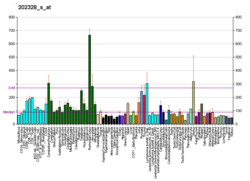
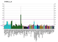
Polycystin 1 (PC1) is a protein that in humans is encoded by the PKD1 gene.[5][6] Mutations of PKD1 are associated with most cases of autosomal dominant polycystic kidney disease, a severe hereditary disorder of the kidneys characterised by the development of renal cysts and severe kidney dysfunction.[7]
Protein structure and function

PC1 is a membrane-bound protein 4303 amino acids in length expressed largely upon the primary cilium, as well as apical membranes, adherens junctions, and desmosomes.[8] It has 11 transmembrane domains, a large extracellular N-terminal domain, and a short (about 200 amino acid) cytoplasmic C-terminal domain.[8][9] This intracellular domain contains a coiled-coil domain through which PC1 interacts with polycystin 2 (PC2), a membrane-bound Ca2+-permeable ion channel.
PC1 has been proposed to act as a G protein–coupled receptor.[8][10] The C-terminal domain may be cleaved in a number of different ways. In one instance, a ~35 kDa portion of the tail has been found to accumulate in the cell nucleus in response to decreased fluid flow in the mouse kidney.[11] In another instance, a 15 kDa fragment may be yielded, interacting with transcriptional activator and co-activator STAT6 and p100, or components of the canonical Wnt signaling pathway in an inhibitory manner.[12][13]
The structure of the human PKD1-PKD2 complex has been solved by cryo-electron microscopy, which showed a 1:3 ratio of PKD1 and PKD2 in the structure. PKD1 consists of a voltage-gated ion channel fold that interacts with PKD2.[14]
PC1 mediates mechanosensation of fluid flow by the primary cilium in the renal epithelium and of mechanical deformation of articular cartilage.[15]
Gene
Splice variants encoding different isoforms have been noted for PKD1. The gene is closely linked to six pseudogenes in a known duplicated region on chromosome 16p.[16]
References
External links
Wikiwand in your browser!
Seamless Wikipedia browsing. On steroids.
Every time you click a link to Wikipedia, Wiktionary or Wikiquote in your browser's search results, it will show the modern Wikiwand interface.
Wikiwand extension is a five stars, simple, with minimum permission required to keep your browsing private, safe and transparent.


