Optic vesicle
Sac that protrudes from the embryonic forebrain to form each eye From Wikipedia, the free encyclopedia

The eyes begin to develop as a pair of diverticula (pouches) from the lateral aspects of the forebrain. These diverticula make their appearance before the closure of the anterior end of the neural tube;[1][2] after the closure of the tube around the 4th week of development, they are known as the optic vesicles. Previous studies of optic vesicles suggest that the surrounding extraocular tissues – the surface ectoderm and extraocular mesenchyme – are necessary for normal eye growth and differentiation.[3]
| Optic vesicle | |
|---|---|
Transverse section of head of chick embryo of forty-eight hours’ incubation. (Optic vesicle labeled at lower right.) | |
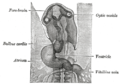
 Human embryo about fifteen days old. Brain and heart represented from right side. Digestive tube and yolk sac in median section. (Optic vesicle labeled at center top.) | |
| Details | |
| Carnegie stage | 11 |
| Gives rise to | Human eyes |
| Identifiers | |
| Latin | vesicula optica; vesicula ophthalmica |
| TE | vesicle_by_E5.14.3.4.2.2.4 E5.14.3.4.2.2.4 |
| Anatomical terminology | |
They project toward the sides of the head, and the peripheral part of each expands to form a hollow bulb, while the proximal part remains narrow and constitutes the optic stalk, which goes on to form the optic nerve.[4][5]
Additional images
 Head of chick embryo of about thirty-eight hours’ incubation, viewed from the ventral surface. X 26

See also
References
External links
Wikiwand - on
Seamless Wikipedia browsing. On steroids.