Top Qs
Timeline
Chat
Perspective
User:Kwyjibo87/Chromosome
From Wikipedia, the free encyclopedia
Remove ads
This is a sandbox for my work on chromosome wiki page
A chromosome is an organized structure of DNA and protein that is found in cells. It is a single, continuous molecule of double-stranded DNA containing many genes, regulatory elements, and other nucleotide sequences. In cells, chromosomal DNA is not "naked"; rather, it is wrapped around DNA-binding proteins known as histones which serve to package the DNA and control its functions. A cell may contain many chromosomes, with the collection of all the chromosomes in a cell known as the genome. The word chromosome comes from the Greek χρῶμα (chroma, color) and σῶμα (soma, body) due to their property of being very strongly stained by particular dyes.

Chromosomes vary widely between different organisms, with the most striking differences observed between prokaryotes (organisms lacking cell nuclei) and eukaryotes (cells containing nuclei). The DNA molecule may be circular or linear, and can be composed of 10,000 to 1,000,000,000[1] nucleotides in a long chain. Typically eukaryotic cells (cells with nuclei) have large linear chromosomes and prokaryotic cells (cells without defined nuclei) have smaller circular chromosomes, although there are many exceptions to this rule. Furthermore, cells may contain more than one type of chromosome; for example, mitochondria in most eukaryotes and chloroplasts in plants have their own small chromosomes.
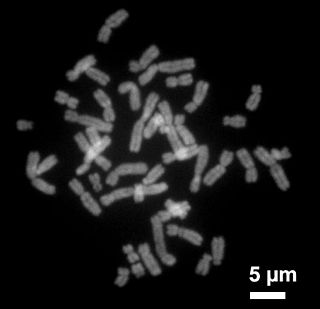
In eukaryotes, nuclear chromosomes are packaged by proteins into a condensed structure called chromatin. This allows the very long DNA molecules to fit into the cell nucleus. The structure of chromosomes and chromatin varies through the cell cycle. Chromosomes are the essential unit for cellular division and must be replicated, divided, and passed successfully to their daughter cells so as to ensure the genetic diversity and survival of their progeny. Chromosomes may exist as either duplicated or unduplicated—unduplicated chromosomes are single linear strands, whereas duplicated chromosomes (copied during synthesis phase) contain two copies joined by a centromere. Compaction of the duplicated chromosomes during mitosis and meiosis results in the classic four-arm structure (pictured to the right). Chromosomal recombination plays a vital role in genetic diversity. If these structures are manipulated incorrectly, through processes known as chromosomal instability and translocation, the cell may undergo mitotic catastrophe and die, or it may aberrantly evade apoptosis leading to the progression of cancer.
In practice "chromosome" is a rather loosely defined term. In prokaryotes and viruses, the term genophore is more appropriate when no chromatin is present. However, a large body of work uses the term chromosome regardless of chromatin content. In prokaryotes DNA is usually arranged as a circle, which is tightly coiled in on itself, sometimes accompanied by one or more smaller, circular DNA molecules called plasmids. These small circular genomes are also found in mitochondria and chloroplasts, reflecting their bacterial origins. The simplest genophores are found in viruses: these DNA or RNA molecules are short linear or circular genophores that often lack structural proteins.
Remove ads
History
Summarize
Perspective
Identification of Chromosomes
The idea that the hereditary information of a cell was contained within the nucleus originated within Ernst Haeckel's Generelle Morphologie of 1866.[2] While insightful, at the time Haeckel's notion lacked supporting scientific data. The first direct observation of chromosomes was achieved in 1879 by Walter Flemming, who used microscopy to observe mitosis (cell division)in salamander tail cells. Using dyes to aid in observing mitosis, he took note of strongly stained thread-like structures he coined chromatin. A colleague of Flemming, Heinrich Wilhelm Gottfried von Waldeyer-Hartz later renamed the stained structures chromosomes.
The evidence for this insight gradually accumulated until, after twenty or so years, two of the greatest in a line of great German scientists[citation needed] spelled out the concept. August Weismann proposed that the germ line is separate from the soma, and that the cell nucleus is the repository of the hereditary material, which, he proposed, is arranged along the chromosomes in a linear manner. Further, he proposed that at fertilisation a new combination of chromosomes (and their hereditary material) would be formed. This was the explanation for the reduction division of meiosis (first described by van Beneden).
Chromosomes as Vectors of Inheritance
In 1902, Theodor Boveri demonstrated through a series of experiments using sea urchin sperm and egg cells the following three properties of chromosomes:
- That the nucleus is the cellular component responsible for heredity
- That chromosomes are likely the vectors of heredity
- That each chromosome contains a unique set of hereditary information necessary for proper cell development
It is worthy to note that the third property Boveri identified was possible due to chromosomes having distinct morphologies under a microscope.
It is the second of these principles that was so original[citation needed]. Boveri was able to test the proposal put forward by Wilhelm Roux, that each chromosome carries a different genetic load, and showed that Roux was right. Upon the rediscovery of Mendel, Boveri was able to point out the connection between the rules of inheritance and the behaviour of the chromosomes. It is interesting to see that Boveri influenced two generations of American cytologists: Edmund Beecher Wilson, Walter Sutton and Theophilus Painter were all influenced by Boveri (Wilson and Painter actually worked with him).
In his famous textbook The Cell, Wilson linked Boveri and Sutton together by the Boveri-Sutton theory. Mayr remarks that the theory was hotly contested by some famous geneticists: William Bateson, Wilhelm Johannsen, Richard Goldschmidt and T.H. Morgan, all of a rather dogmatic turn-of-mind. Eventually complete proof came from chromosome maps in Morgan's own lab.[3]
Remove ads
Chromosomes in Prokaryotes
Summarize
Perspective
The prokaryotes – bacteria and archaea – typically have a single circular chromosome, but many variations do exist.[4] Most bacteria have a single circular chromosome that can range in size from only 160,000 base pairs in the endosymbiotic bacterium Candidatus Carsonella ruddii,[5] to 12,200,000 base pairs in the soil-dwelling bacterium Sorangium cellulosum.[6] Spirochaetes of the genus Borrelia are a notable exception to this arrangement, with bacteria such as Borrelia burgdorferi, the cause of Lyme disease, containing a single linear chromosome.[7]
Structure in sequences
Prokaryotic chromosomes have less sequence-based structure than eukaryotes. Bacteria typically have a single point (the origin of replication) from which replication starts, whereas some archaea contain multiple replication origins.[8] The genes in prokaryotes are often organized in operons, and do not usually contain introns, unlike eukaryotes.
DNA packaging
Prokaryotes do not possess nuclei. Instead, their DNA is organized into a structure called the nucleoid.[9] The nucleoid is a distinct structure and occupies a defined region of the bacterial cell. This structure is, however, dynamic and is maintained and remodeled by the actions of a range of histone-like proteins, which associate with the bacterial chromosome.[10] In archaea, the DNA in chromosomes is even more organized, with the DNA packaged within structures similar to eukaryotic nucleosomes.[11][12]
Bacterial chromosomes tend to be tethered to the plasma membrane of the bacteria. In molecular biology application, this allows for its isolation from plasmid DNA by centrifugation of lysed bacteria and pelleting of the membranes (and the attached DNA).
Prokaryotic chromosomes and plasmids are, like eukaryotic DNA, generally supercoiled. The DNA must first be released into its relaxed state for access for transcription, regulation, and replication.
Remove ads
Chromosomes in Eukaryotes
Summarize
Perspective
(tag removed) mergefrom|Eukaryotic chromosome fine structure|date=December 2007}} Eukaryotes (cells with nuclei such as those found in plants, yeast, and animals) possess multiple large linear chromosomes contained in the cell's nucleus. Each chromosome has one centromere, with one or two arms projecting from the centromere, although, under most circumstances, these arms are not visible as such. In addition, most eukaryotes have a small circular mitochondrial genome, and some eukaryotes may have additional small circular or linear cytoplasmic chromosomes. The ends of the linear chromosome arms contain highly condensed, repetitive DNA sequences known as telomeres.
In the nuclear chromosomes of eukaryotes, the uncondensed or "naked" DNA is packaged by being wrapped around globular proteins called histones (structural proteins), forming a composite structure known as a nucleosome. Each nucleosome consists 147 base-pairs of double-stranded DNA wrapped around a single histone protein. The general term for DNA bound by packaging proteins is chromatin.
Chromatin
Chromatin is the complex of DNA and protein found in the eukaryotic nucleus, which packages chromosomes. The structure of chromatin varies significantly between different stages of the cell cycle, according to the requirements of the DNA. Chromatin should be distinguished from DNA which is "naked", or unpackaged by DNA-packaging proteins such as histones.
Chromatin in eukaryotic cells can be found in two general states:
- Euchromatin, which consists of DNA that is loosely packaged and tends to contain actively expressed genes.
- Heterochromatin, which consists of mostly inactive (non-expressed) DNA and is more highly compacted than euchromatin. Heterochromatin can be further distinguished into two types:
- Constitutive heterochromatin, which is never expressed. It is located around the centromere and usually contains repetitive sequences. Constituitive heterochromatin is also associated with silencing of mobile DNA elements such as transposons.
- Facultative heterochromatin, which is sometimes expressed. Facultative heterochromatin is often used to regulate the expression of genes in differentiated cells (for example, a muscle cell would prevent expression of genes which regulate neuron-specific processes).
Individual chromosomes cannot be visually distinguished in interphase without the use of dyes or molecular labels – they appear in the nucleus as a homogeneous tangled mix of DNA and protein.
Metaphase chromatin and division

In the early stages of mitosis or meiosis (cell division), the chromatin strands become more and more condensed. They cease to function as accessible genetic material (transcription stops) and become a compact transportable form. This compact form makes the individual chromosomes visible, and they form the classic four arm structure, a pair of sister chromatids attached to each other at the centromere. The shorter arms are called p arms (from the French petit, small) and the longer arms are called q arms (q follows p in the Latin alphabet). This is the only natural context in which individual chromosomes are visible with an optical microscope.
During divisions, long microtubules attach to the centromere and the two opposite ends of the cell. The microtubules then pull the chromatids apart, so that each daughter cell inherits one set of chromatids. Once the cells have divided, the chromatids are uncoiled and can function again as chromatin. In spite of their appearance, chromosomes are structurally highly condensed, which enables these giant DNA structures to be contained within a cell nucleus (Fig. 2).
The self-assembled microtubules form the spindle, which attaches to chromosomes at specialized structures called kinetochores, one of which is present on each sister chromatid. A special DNA base sequence in the region of the kinetochores provides, along with special proteins, longer-lasting attachment in this region.

Remove ads
Behavior in Cell Cycle
Mitosis
Meiosis
= Homologous Recombination
Sex Determination
Worms XX/X0
Insects XX/XY but determined by amount of X
Birds ZZ/ZW
Mammals XX/XY determined by Y (SRY)
X-inactivation
Genomes
Ploidy
C-value Paradox
Human Genetics
Human Genome
Diseases
Relationship to Cancer
Number of chromosomes in various organisms
Wikiwand - on
Seamless Wikipedia browsing. On steroids.
Remove ads