File:MultiPhotonExcitation-Fig10-doi10.1186slash1475-925X-5-36-clipping.JPEG
From Wikipedia, the free encyclopedia
MultiPhotonExcitation-Fig10-doi10.1186slash1475-925X-5-36-clipping.JPEG (714 × 467 pixels, file size: 81 KB, MIME type: image/jpeg)
| This is a file from the Wikimedia Commons. Information from its description page there is shown below. Commons is a freely licensed media file repository. You can help. |
Summary
| DescriptionMultiPhotonExcitation-Fig10-doi10.1186slash1475-925X-5-36-clipping.JPEG |
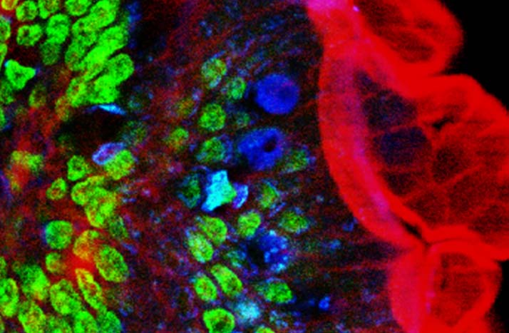
English: Original figure legend: Multiple fluorescence 2PE imaging. 2PE multiple fluorescence image from a 16 μm cryostat section of mouse intestine stained with a combination of fluorescent stains (F-24631, Molecular Probes). Alexa Fluor 350 wheat germ agglutinin, a blue-fluorescent lectin, was used to stain the mucus of goblet cells. The filamentous actin prevalent in the brush border was stained with red-fluorescent Alexa Flu or 568 phalloidin. Finally, the nuclei were stained with SYTOX ® Green nucleic acid stain. Imaging has been performed at 780 nm, 100 x 1.4 NA Leica objective, using a Chameleon XR ultrafast Ti-Sapphire laser (Coherent Inc., USA) coupled at LAMBS-MicroScoBio with a Spectral Confocal Laser Scanning Microscope, Leica SP2-AOBS.
Deutsch: Zweiphotonenaufnahme an einem Schnitt durch einen Mausdarm. Zellkerne in grün, Schleim der Becherzellen in blau, Aktin (Phalloidin-Färbung) in rot. Anregung erfolgte bei 780 nm durch einen Titan:Saphir-Laser.
Français : légende originale de l'image : imagerie en fluorescenc emultiple 2PE d'une section de 16 µm de cryostat d'intestin de souris coloré avec une combinaison de colorants fluorescents (F-24631, Molecular Probes). l'Alexa Fluor 350 d'agglutinine degerme de blé, une lectine bleu fluorescente, a été utilisée pour colorer le mucus des cellules caliciformes. L'actine filamenteuse a été colorée avec du rouge fluorescent (Alexa Flu ou phalloïdine 568). Enfin, les noyaux ont été colorés avec un autre colorant (SYTOX ® Green nucleic acid stain). L'image a été faite à 780 nm, avec un objectif Leica 100 x 1,4 NA, en utilisant un éclairage laser (Chameleon XR ultrafast Ti-Sapphire laser (Coherent Inc., USA) ) couplé à un microscope LAMBS-MicroScoBio (Spectral Confocal Laser Scanning Microscope, Leica SP2-AOBS). |
| Date | Original version: 6 June 2006. Clipping: 4. March 2009. |
| Source |
Multi-photon excitation microscopy. BioMedical Engineering OnLine, 2006, 5:36. |
| Author |
Alberto Diaspro, Paolo Bianchini, Giuseppe Vicidomini, Mario Faretta, Paola Ramoino and Cesare Usai. |
| Permission (Reusing this file) |
This file is licensed under the Creative Commons Attribution 2.0 Generic license.
|
| Other versions | For unclipped version see below |
All images uploaded from this article about multi-photon and two-photon-microscopy:


Captions
Items portrayed in this file
depicts
image/jpeg
File history
Click on a date/time to view the file as it appeared at that time.
| Date/Time | Thumbnail | Dimensions | User | Comment | |
|---|---|---|---|---|---|
| current | 20:57, 4 March 2009 | | 714 × 467 (81 KB) | Dietzel65 | == Beschreibung == {{Information |Description={{en|1=Original figure legend: ''Multiple fluorescence 2PE imaging. 2PE multiple fluorescence image from a 16 μm cryostat section of mouse intestine stained with a combination of fluorescent stains (F-24631, |
File usage
Global file usage
The following other wikis use this file:
- Usage on ar.wikipedia.org
- Usage on ca.wikipedia.org
- Usage on de.wikipedia.org
- Usage on es.wikipedia.org
- Usage on fr.wikipedia.org
- Usage on it.wikipedia.org
- Usage on outreach.wikimedia.org
- Usage on uk.wikipedia.org
- Usage on zh.wikipedia.org
Metadata
This file contains additional information, probably added from the digital camera or scanner used to create or digitize it.
If the file has been modified from its original state, some details may not fully reflect the modified file.
| Author | TCS User |
|---|---|
| Orientation | Normal |
| Horizontal resolution | 72 dpi |
| Vertical resolution | 72 dpi |
| Software used | Adobe Photoshop 7.0 |
| File change date and time | 21:47, 4 March 2009 |
| Y and C positioning | Centered |
| Exif version | 2.2 |
| Color space | Uncalibrated |