File:Anatomy_of_Human_Ear_with_Cochlear_Frequency_Mapping.svg
From Wikipedia, the free encyclopedia

Size of this PNG preview of this SVG file: 674 × 519 pixels. Other resolutions: 312 × 240 pixels | 624 × 480 pixels | 998 × 768 pixels | 1,280 × 986 pixels | 2,560 × 1,971 pixels.
Original file (SVG file, nominally 674 × 519 pixels, file size: 33 KB)
| This is a file from the Wikimedia Commons. Information from its description page there is shown below. Commons is a freely licensed media file repository. You can help. |
Summary
| DescriptionAnatomy of Human Ear with Cochlear Frequency Mapping.svg |
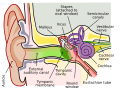
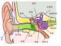
English: The human ear and frequency mapping in the cochlea. The three ossicles incus, malleus, and stapes transmit airborne vibration from the tympanic membrane to the oval window at the base of the cochlea. Because of the mechanical properties of the basilar membrane within the snail-shaped cochlea, high frequencies will produce a vibration peak near the oval window, whereas low frequencies will stimulate receptors near the apex of the cochlea (locations for three frequencies indicated schematically). Information from the cochlear receptor cells is transmitted to the cochlear nuclei via the 8th cranial nerve, and on through the midbrain to the cortex. |
| Date | |
| Source | Own work (Original text: Own work by uploader, derived from File:Anatomy_of_the_Human_Ear.svg) |
| Author | Inductiveload |
| Permission (Reusing this file) |
This file is licensed under the Creative Commons Attribution-Share Alike 2.5 Generic license.
|
| Other versions |
[edit]
|
| SVG development InfoField | This vector image was created with Inkscape. This file is translated using SVG switch elements: all translations are stored in the same file. |


Captions
A labelled cross-sectional diagram of the human ear.
A labelled cross-sectional diagram of the human ear.
Items portrayed in this file
depicts
15 February 2009
image/svg+xml
4c58b13c29ef1eb6b3ba8c18aed2cd55519d51d1
34,114 byte
519 pixel
674 pixel
File history
Click on a date/time to view the file as it appeared at that time.
| Date/Time | Thumbnail | Dimensions | User | Comment | |
|---|---|---|---|---|---|
| current | 21:29, 16 September 2018 | | 674 × 519 (33 KB) | JoKalliauer | added systemLanguage="eo" |
| 17:21, 16 September 2018 |  | 674 × 519 (32 KB) | JoKalliauer | added systemLanguage="de" | |
| 05:33, 11 September 2018 |  | 674 × 519 (87 KB) | Jmarchn | Bigger (proportional real size) and full redraw (more realistic) of the auricle. Ossicles in white colour. Eardrum with contour. Added 3 labels. Add fundus to the bone and subcutaneous tissues, add superior auricular muscle, add transparency to middle ear, add separation between middle and inner ear, add division to internal auditory canal. | |
| 13:40, 29 April 2009 |  | 800 × 600 (98 KB) | Inductiveload | swap incus/malleus | |
| 15:10, 15 February 2009 |  | 800 × 600 (98 KB) | Inductiveload | {{Information |Description={{en|1=The human ear and frequency mapping in the cochlea. The three ossicles incus, malleus, and stapes transmit airborne vibration from the tympanic membrane to the oval window at the base of the cochlea. Because of the mechan |
File usage
The following pages on the English Wikipedia use this file (pages on other projects are not listed):
Global file usage
The following other wikis use this file:
- Usage on en.wikibooks.org
- Usage on eo.wikipedia.org
- Usage on he.wikipedia.org
- Usage on lt.wikipedia.org
- Usage on meta.wikimedia.org
- Usage on www.wikidata.org
Metadata
This file contains additional information, probably added from the digital camera or scanner used to create or digitize it.
If the file has been modified from its original state, some details may not fully reflect the modified file.
| Width | 674.23 |
|---|---|
| Height | 518.84 |