File:Thyroid_papillary_carcinoma_histopathology_(4).jpg
From Wikipedia, the free encyclopedia
No higher resolution available.
Thyroid_papillary_carcinoma_histopathology_(4).jpg (500 × 376 pixels, file size: 59 KB, MIME type: image/jpeg)
| This is a file from the Wikimedia Commons. Information from its description page there is shown below. Commons is a freely licensed media file repository. You can help. |
Summary
| DescriptionThyroid papillary carcinoma histopathology (4).jpg |
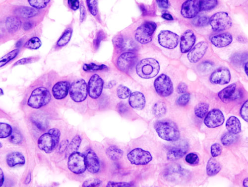
English: Histopatholgical image of papillary carcinoma of the thyroid gland obtained by a total thyroidectomy. Hematoxylin and eosin stain. Another version of an accompanying file "Thyroid_papillary_carcinoma_histopathology_(3).jpg". |
| Source | No machine-readable source provided. Own work assumed (based on copyright claims). |
| Author | No machine-readable author provided. KGH assumed (based on copyright claims). |
Licensing
I, the copyright holder of this work, hereby publish it under the following licenses:

|
Permission is granted to copy, distribute and/or modify this document under the terms of the GNU Free Documentation License, Version 1.2 or any later version published by the Free Software Foundation; with no Invariant Sections, no Front-Cover Texts, and no Back-Cover Texts. A copy of the license is included in the section entitled GNU Free Documentation License.http://www.gnu.org/copyleft/fdl.htmlGFDLGNU Free Documentation Licensetruetrue |
| This file is licensed under the Creative Commons Attribution-Share Alike 3.0 Unported license. | ||
| ||
| This licensing tag was added to this file as part of the GFDL licensing update.http://creativecommons.org/licenses/by-sa/3.0/CC BY-SA 3.0Creative Commons Attribution-Share Alike 3.0truetrue |
You may select the license of your choice.
| Annotations InfoField | This image is annotated: View the annotations at Commons |
282
126
{{{4}}}
31
500
376
щи# Nuclear pseudoinclusion
Captions
Histopatholgical image of papillary carcinoma of the thyroid gland
Items portrayed in this file
depicts
File history
Click on a date/time to view the file as it appeared at that time.
| Date/Time | Thumbnail | Dimensions | User | Comment | |
|---|---|---|---|---|---|
| current | 14:55, 8 January 2006 | | 500 × 376 (59 KB) | KGH | Histopatholgical image of papillary carcinoma of the thyroid gland obtained by a total thyroidectomy. Hematoxylin and eosin stain. Another version of an accompanying file "Thyroid_papillary_carcinoma_histopathology_(3).jpg". |
File usage
The following pages on the English Wikipedia use this file (pages on other projects are not listed):
Global file usage
The following other wikis use this file:
- Usage on ar.wikipedia.org
- Usage on bn.wikipedia.org
- Usage on bs.wikipedia.org
- Usage on ca.wikipedia.org
- Usage on es.wikipedia.org
- Usage on fa.wikipedia.org
- Usage on gl.wikipedia.org
- Usage on hy.wikipedia.org
- Usage on it.wikipedia.org
- Usage on ja.wikipedia.org
- Usage on ko.wikipedia.org
- Usage on new.wikipedia.org
- Usage on or.wikipedia.org
- Usage on pt.wikipedia.org
- Usage on ru.wikipedia.org
- Usage on sh.wikipedia.org
- Usage on sk.wikipedia.org
- Usage on sr.wikipedia.org
- Usage on sw.wikipedia.org
- Usage on tr.wikipedia.org
- Usage on zh.wikipedia.org
Metadata
This file contains additional information, probably added from the digital camera or scanner used to create or digitize it.
If the file has been modified from its original state, some details may not fully reflect the modified file.
| Image title | OLYMPUS DIGITAL CAMERA |
|---|---|
| Camera manufacturer | OLYMPUS OPTICAL CO.,LTD. |
| Camera model | DP70 |
| Exposure time | 2/11 sec (0.18181818181818) |
| ISO speed rating | 200 |
| Date and time of data generation | 2005/07/06 13:26:06 |
| Horizontal resolution | 72 dpi |
| Vertical resolution | 72 dpi |
| File change date and time | 2005/07/06 13:26:06 |
| Y and C positioning | Co-sited |
| Exif version | 2 |
| Date and time of digitizing | 2005/07/06 13:26:16 |
| Shutter speed | 2.959 |
| Metering mode | Spot |
| Light source | Unknown |
| Color space | sRGB |
| Sensing method | One-chip color area sensor |
.jpg)