User:Jakob Suckale/panel bottom
From Wikipedia, the free encyclopedia
!
Diagrams & Photos contributed
|- |
 Amino acid composition in food and blood 11/16
 Mouse pancreatic islet 9/11
 Sep 2008 for acyclovir


 9/07 illustration explaining h-index
 5/07 amylin (IAPP) structure
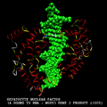 5/07 HNF1a bound to DNA; from BioInfo Bank with permission
 5/07 structure of protein HNF1a bound to DNA, PDB 1IC8
 5/07 structure of protein TCF7L2, PDB 2GL7 made with KiNG
 4/07 Hygromycin B structure copied from PubChem
 3/07 logo via screenshot of PLoSone.org, CC2.5
 3/07 logo via screenshot of PLoS.org, CC2.5
 8/06 schematic of protein sorting signals
 5/06 visualisation of the ancient Greek four humours
 5/06 structure of Tris
- 5/06 structure of Cresol Red
- 5/06 photo of MPI-CBG by K. Margitudis uploaded
 5/06 simple diagram representing the idea of flora made
 5/06 simple diagram representing the idea of fauna made
 5/06 bio-barnstar suggestion 4 cropped & retouched
 5/06 bio-barnstar suggestion 3 cropped & retouched
 5/06 bio-barnstar suggestion 2 cropped & retouched; idea by LiquidGhoul
 5/06 bio-barnstar suggestion composed from various photos
 3/06 hSUMO structure diagram made with iMol
 3/06 hSUMO structure diagram made with iMol
 11/05 screenshot uploaded


_for_wikipedia_article_of_the_same_name.png)


- Some fair use logos I uploaded but which cannot be displayed here:
|-