File:Loupe-binoculaire-p1030891.jpg
From Wikipedia, the free encyclopedia
Size of this preview: 800 × 594 pixels. Other resolutions: 320 × 238 pixels | 640 × 475 pixels | 1,024 × 760 pixels | 1,280 × 950 pixels | 2,535 × 1,882 pixels.
Original file (2,535 × 1,882 pixels, file size: 2.72 MB, MIME type: image/jpeg)
| This is a file from the Wikimedia Commons. Information from its description page there is shown below. Commons is a freely licensed media file repository. You can help. |
Summary
| DescriptionLoupe-binoculaire-p1030891.jpg |
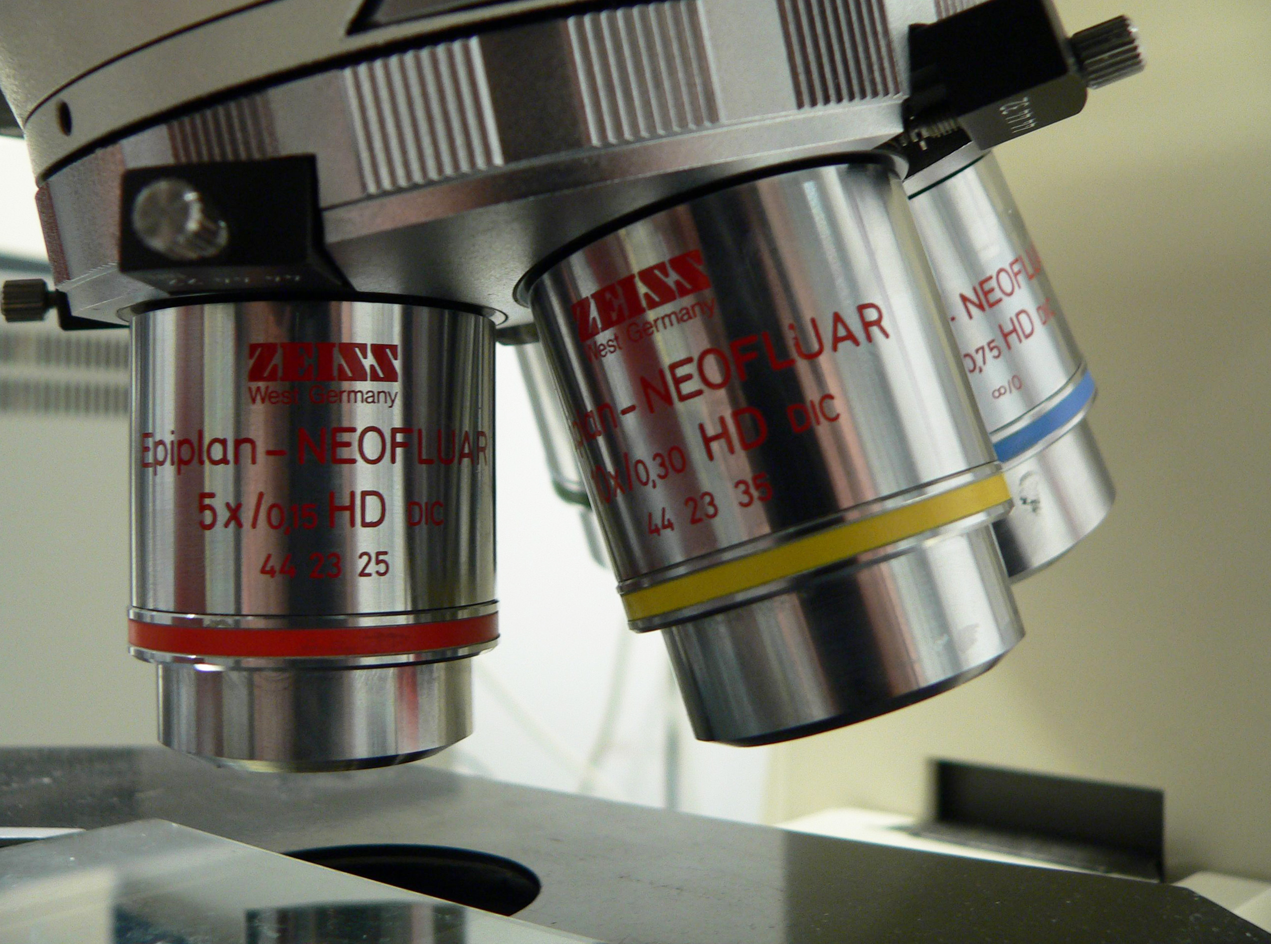
English: binocular microscope
Français : Loupe binoculaire |
||
| Date |
Unknown date Unknown date |
||
| Source | Own work | ||
| Author | Rama | ||
| Permission (Reusing this file) |
This file is licensed under the Creative Commons Attribution-Share Alike 2.0 France license.
 |

Captions
Add a one-line explanation of what this file represents
Items portrayed in this file
depicts
File history
Click on a date/time to view the file as it appeared at that time.
| Date/Time | Thumbnail | Dimensions | User | Comment | |
|---|---|---|---|---|---|
| current | 15:13, 19 December 2015 | | 2,535 × 1,882 (2.72 MB) | Jacek Halicki | tilt, more light, jpg compression |
| 13:43, 17 March 2006 |  | 2,560 × 1,920 (546 KB) | Rama | {{fr|Loupe binoculaire}} {{en|binocular microscope}} {{Rama}} Category:Microscopes |
File usage
The following pages on the English Wikipedia use this file (pages on other projects are not listed):
- 4Pi microscope
- American Microscopical Society
- Antonie van Leeuwenhoek
- Bright-field microscopy
- Confocal microscopy
- Critical illumination
- Dark-field microscopy
- Differential interference contrast microscopy
- Diffraction-limited system
- Dispersion staining
- Fluorescence microscope
- Köhler illumination
- Lattice light-sheet microscopy
- Light sheet fluorescence microscopy
- Live-cell imaging
- Microscope
- Microscopy
- Near-field scanning optical microscope
- Objective (optics)
- Optical microscope
- Optical sectioning
- Phase-contrast microscopy
- Phase telescope
- Photoactivated localization microscopy
- Quantitative phase-contrast microscopy
- Raman microscope
- STED microscopy
- Sarfus
- Second-harmonic imaging microscopy
- Super-resolution microscopy
- Superlens
- Three-photon microscopy
- Time-lapse microscopy
- Total internal reflection fluorescence microscope
- Two-photon excitation microscopy
- Vertico spatially modulated illumination
- Talk:Microscope/Archive 1
- User:Egelberg/sandbox
- User:Lamals/sandbox/Fluorescence microscopy via coherent control
- User:Lamals/sandbox/STED microscopy
- User:Telementor/Userboxes/microscopy
- Template:Optical microscopy
Global file usage
The following other wikis use this file:
- Usage on af.wikipedia.org
- Usage on ar.wikipedia.org
- Usage on bn.wikipedia.org
- Usage on es.wikipedia.org
- Usage on fa.wikipedia.org
- Usage on fr.wikipedia.org
- Usage on he.wikipedia.org
- Usage on hr.wikipedia.org
- Usage on ja.wikipedia.org
- Usage on ko.wikipedia.org
- Usage on nl.wikipedia.org
- Usage on no.wikipedia.org
- Usage on pt.wikipedia.org
- Usage on sr.wikipedia.org
- Usage on szy.wikipedia.org
- Usage on th.wikipedia.org
- Usage on tr.wikipedia.org
- Usage on uk.wikipedia.org
- Usage on zh.wikipedia.org
View more global usage of this file.
Metadata
This file contains additional information, probably added from the digital camera or scanner used to create or digitize it.
If the file has been modified from its original state, some details may not fully reflect the modified file.
| Width | 2,560 px |
|---|---|
| Height | 1,920 px |
| Bits per component |
|
| Pixel composition | RGB |
| Orientation | Normal |
| Number of components | 3 |
| Horizontal resolution | 72 dpi |
| Vertical resolution | 72 dpi |
| Software used | Adobe Photoshop CS6 (Windows) |
| File change date and time | 16:12, 19 December 2015 |
| Exif version | 2.21 |
| Color space | Uncalibrated |
| Unique ID of original document | 050FFA8E59A2C7999CFF913C29E5C562 |
| Date and time of digitizing | 17:09, 19 December 2015 |
| Date metadata was last modified | 17:12, 19 December 2015 |
Retrieved from "https://en.wikipedia.org/wiki/File:Loupe-binoculaire-p1030891.jpg"